The spinal cord cross section unlabeled invites us on an intriguing journey into the intricacies of the central nervous system. This comprehensive guide delves into the anatomical architecture of the spinal cord, unraveling the complexities of its gray and white matter, the protective layers of meninges, and the intricate network of blood vessels that sustain its vitality.
Through meticulous descriptions and insightful explanations, we will illuminate the functional significance of each component, gaining a profound understanding of this vital conduit of communication and control.
Overview of Spinal Cord Cross Section: Spinal Cord Cross Section Unlabeled

The spinal cord is a long, cylindrical structure that extends from the brainstem to the lumbar region. It is enclosed within the vertebral canal of the spine and is responsible for transmitting sensory and motor information between the brain and the rest of the body.
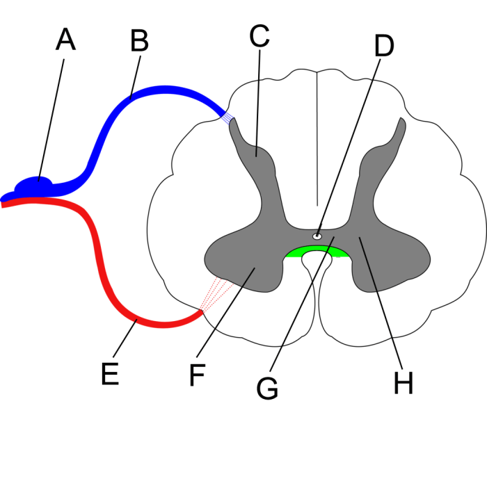
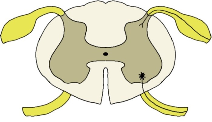
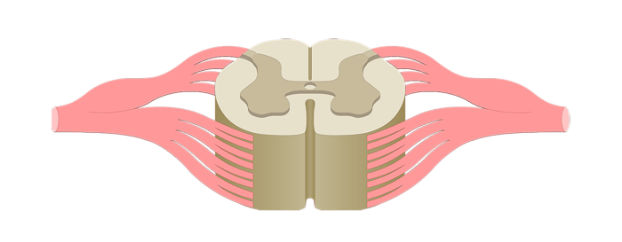
A cross section of the spinal cord reveals a complex structure consisting of gray matter and white matter. The gray matter, located in the center of the spinal cord, contains the cell bodies of neurons, while the white matter, located on the periphery, contains the axons of neurons.
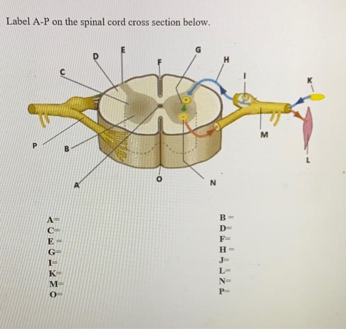
A labeled diagram of a spinal cord cross section can help visualize the different structures and their relationships.
Gray Matter of the Spinal Cord, Spinal cord cross section unlabeled
The gray matter of the spinal cord is located in the center of the spinal cord and is shaped like an “H” or a butterfly. It contains the cell bodies of neurons, which are the primary functional units of the nervous system.
There are three main types of neurons found in the gray matter: sensory neurons, motor neurons, and interneurons.
Sensory neurons receive sensory information from the body and transmit it to the brain. Motor neurons transmit motor commands from the brain to the muscles and glands. Interneurons connect sensory and motor neurons within the spinal cord, allowing for complex reflex actions.
The gray matter is divided into several regions, including the dorsal horn, ventral horn, and lateral horn. Each region contains different types of neurons and performs specific functions.
White Matter of the Spinal Cord
The white matter of the spinal cord is located on the periphery of the spinal cord and surrounds the gray matter. It contains the axons of neurons, which are long, slender extensions that transmit electrical signals. The axons are myelinated, which means they are covered in a fatty substance called myelin.
Myelin acts as an insulator, allowing electrical signals to travel quickly and efficiently.
The white matter is divided into three main columns: the dorsal column, lateral column, and ventral column. Each column contains different types of fibers that transmit specific types of information.
The white matter is responsible for transmitting sensory and motor information between the brain and the rest of the body.
Q&A
What is the significance of the gray matter in the spinal cord?
The gray matter, located in the center of the spinal cord, contains the cell bodies of neurons and is responsible for processing and integrating sensory and motor information.
What are the different types of fibers found in the white matter?
The white matter, located in the outer region of the spinal cord, contains myelinated axons that transmit sensory and motor signals to and from the brain.
What is the function of the meninges?
The meninges are three layers of protective membranes that surround the spinal cord, providing structural support, cushioning, and protection from infection.


